Namibia Rhino Study

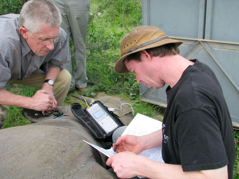
Drs. Robin Radcliffe (right) and Robin Gleed (left) measure the end expired carbon dioxide gas in an anesthetized black rhinoceros. Award funded by the Morris Animal Foundation (Grant # D09ZO-012) in partnership with Cornell University College of Veterinary Medicine and the Namibia Ministry of Environment and Tourism.
Collaboration
Dr. Robin Radcliffe of the Cornell Conservation Medicine Program led a team of scientists to Namibia for a two-year Morris Animal Foundation-funded project to study the respiratory and thermoregulatory patterns of black rhinoceros during field capture. This work was made possible through a collaborative effort with the Namibian Ministry of Environment and Tourism’s Rhino Capture Unit under the direction of Pierre Du Preez and Mark Jago with support from rhinoceros expert, Pete Morkel.
Project Summary
Etosha National Park spans the northcentral and northwestern portion of Namibia and encompasses a diverse landscape of mixed acacia thorn scrub, open plains and mopane woodland, all of which surrounds the vast Etosha pan. This oasis in an otherwise arid land abounds with wildlife including lions and leopard, giraffe and zebra, oryz and gazelle, and both black and white rhinoceros. Here in the jewel of Namibia lies one of Africa’s largest populations of the endangered desert black rhinoceros – Diceros bicornis bicornis. It was here that we came to study and learn about the rhinoceros. Capture and anesthesia of rhinoceros across Africa has been practiced for half a century and the principles of field anesthesia have been well established. Yet for all of this pioneering work there remains a dearth of scientifically based information on the most fundamental aspects of the anesthesia process in these prehistoric beasts. Our team was a rare mix of wildlife veterinarians (Robin Radcliffe and Michelle Miller), University veterinary medical professor (Robin Gleed) and University human medical professor (Art Taft). Our team, funded by the Morris Animal Foundation and partnering with Cornell University and the Ministry of Environment and Tourism, traveled to Namibia to begin a multiyear investigation looking into some of the most challenging aspects of rhinoceros anesthesia: How much air does a rhinoceros breath? What volume of air is moved in and out and what portion of that volume is contributing to gas exchange? How does ventilation and perfusion change with posture? These are a few of the many questions the team set out to answer.
 Over the two-year period the Ministry of Environment and Tourism Game Capture Unit immobilized fifty-four black rhinoceros for routine ear-notching and radiotelemetry monitoring; data collection was conducted opportunistically. Blood samples were collected at regular intervals to measure blood gas and chemistry values in free-ranging animals under anesthesia. At the same time, in a subset of animals, the team collected the first in depth information on core ventilation parameters including tidal volume, minute ventilation, and dead space. These measurements will help rhino scientists make informed decisions about the effects of potent opioid anesthetic drugs and how posture may alter the respiratory and cardiovascular systems; such knowledge may help devise better ways to manage rhinoceros during capture and anesthesia in both the field and zoological setting.
Despite the many challenges of keeping pace with a game capture unit in the wilds of the African bush, the investigation went remarkably well. The gas collection apparatus designed and built by Dr. Robin Gleed worked extremely well and proved suitable for studying how a wild rhinoceros breathes after capture and in various body positions. The apparatus, consisting of large four-inch PVC pipes in the shape of giant candy-cane, was soon dubbed the “hamster run” because Pierre Du Preez pictured his son fancying such a system of pipes for his pet hamsters. The gas collection apparatus was simple in that it had no moving parts and no electronics, but its function was no less impressive. The simple design of pipes separated by a series of one-way valves allowed the investigators to completely separate the inspired air from expired air. In this way, the team was able to collect and measure the entire volume of air that a rhino breathes over a minute (minute ventilation). This basic information is helping to shed new light on the effects of the capture process on rhinoceros. How do rhinos cope with gas exchange in times of increased demand at capture? Does body positioning (sternal compared to lateral recumbency) affect the rhino’s capacity to recover from anesthesia? What effect does pregnancy have on respiratory function and anesthesia outcomes? These are a few of the many questions the investigators are exploring with the help of the Morris Animal Foundation. The information gathered will help scientists determine the precise breathing patterns of recumbent anesthetized rhinoceros and may lead to improved capture and handling protocols for rhinoceros that are routinely moved across Africa to fulfill rigorous conservation strategies for protection of these endangered megavertebrates.